AI-Powered Radiology Interpretation
Experience the power of advanced artificial intelligence for accurate, detailed reports for X-rays, MRIs, CT scans, and more. Swift, reliable, and easy to understand.
One report, two readings. Plain language and clinical terms in the same document — download as PDF or DOCX.
Questions after reading it? Ask them in a private chat attached to your own report — free account required.
Human and veterinary scans alike. Species-appropriate terms, not human ones applied to an animal.
Upload any imaging file
Capabilities
Comprehensive Imaging Analysis
Quick & Reliable
Get accurate radiology interpretations in seconds. No more waiting days for results.
Easy to Understand
We translate complex medical jargon into simple, clear language for patients and pet owners.
Human & Veterinary
Our AI is trained to interpret scans for both humans and their furry companions.
Scan Series Comparison
Compare scan results from the same patient across different dates to track treatment progress and detect changes easily.
Powered by
Process
How It Works
Get professional-grade insights in three simple steps. Designed for speed, accuracy, and clarity.
Upload Your Image
Quickly upload your scan through our secure and encrypted platform. We support X-rays, MRIs, and CT scans.
Receive Your Report
Get a detailed, AI-generated report within seconds. We provide clinical findings alongside easy-to-understand summaries.
Ask Follow-Up Questions
Use our interactive chat assistant to ask questions and get further explanations about your specific results.
This is what your report
will look like
Full AI radiology report — every section, every detail, with plain-language explanations alongside the clinical text. Try different scan types:
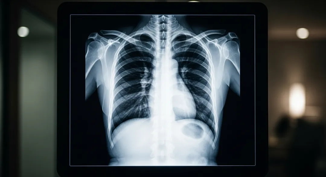 RChest X-Ray · PA · AI-generated image
This is a chest X-ray, PA view. The lungs are mostly clear with mild increased markings in the left lower area. Heart within normal size. The AI identified 4 specific observations in the full report.
PA chest radiograph of diagnostic quality. No prior studies available for comparison.
Your first full report is free
Create a free account — upload your scan and get a report just like this one.
What it can do, and what it can't
- Describes your scan in plain language in under a minute. It runs on Google's Gemini model.
- For a DICOM series, it reviews 9–10 slices selected at even intervals.
- To be direct: the model is general-purpose, without medical certification.
- No doctor checks the result — that's why it costs a few dollars, not two hundred.
Independent research on AI in radiology
This research concerns AI radiology technology in general, not this product. Links open the original publications.
Ready to upload your scan?
Your first full report is free. No credit card required.
Not an X-ray?
Other scan types arrive as different files and use different words. Each has its own page.
CT Scan Report Analysis Online
A few hundred cross-sections, reconstructed several ways in the same folder.
Open the CT page →Free AI MRI Report Analysis
Several sequences per exam — T1, T2, FLAIR, DWI — each describing the same tissue differently.
Open the MRI page →Free AI Ultrasound Report Reader
Usually a handful of still frames captured from a live, operator-driven exam.
Open the ultrasound page →Knowledge Hub
Knowledge Hub Insights
Explore our latest tutorials, industry news, and clinical insights to get the most out of your AI radiology experience.
Stay Updated
Get the latest AI radiology insights and platform updates delivered directly to your inbox.
Trusted by users worldwide
NPS 84 · 88% rate us 9–10Feedback collected after real scan analyses
“The report included the specific red flag areas on a dental x-ray and possible factors causing them […] It also allows for follow-up questions to the AI Assistant”
Verified user · Jun 2026
“this report was so fast and picked up every finding I had identified […]”
Verified user · Apr 2026
“Me ha sido muy mucha ayuda gracias”
Verified user · May 2026
Based on 25 post-analysis ratings collected Feb–Jul 2026
Our Mission
Democratizing Radiology with AI
We strive to bridge the gap between medical imaging and immediate, actionable insights. By combining state-of-the-art artificial intelligence with clinical expertise, we aim to reduce diagnostic wait times, support medical professionals, and empower patients with clarity and understanding.
Analysis in seconds, not days
Accessible from anywhere
Clinical-grade reasoning
About Us
Pioneering the Future of Diagnostic Imaging
Founded by a coalition of AI Vision Consultants and radiologists, X-ray AI Analyzer leverages AI models to provide instant and easy to understand Analyzes to patients, as well as second-opinion radiology reports.
Our platform ensures that advanced medical imaging analysis is accessible not just to large hospitals, but to remote clinics, urgent care centers, and veterinary practices around the globe.
"The anxiety of not understanding my MRI report was overwhelming. This tool translated the MRI into plain language instantly, giving me peace of mind before seeing my specialist."