MRI scan analysis
Understand Your MRI Images with AI
Upload images from your MRI — a DICOM folder from the hospital disc or exported JPG/PNG files. The AI explains what the selected images show in plain language, next to a clinical-style report. Your first analysis is free with a free account, and you can ask follow-up questions about the report in a chat.
This page analyses images. If you have the written MRI report, add it (PDF or photo) as optional context next to the images; a report on its own is not analysed.
How it works
Choose your study filesDrop the MRI folder or select files: DICOM (.dcm, DICOMDIR, files without an extension) or JPG/PNG images.
Pick the images and add contextChoose up to 10 images from the sequences you want explained, enter the patient's age and, if you like, symptoms, notes or the written report.
Get the explanation and ask questionsAfter a free sign-in the analysis runs and you get the report. Ask follow-up questions in the chat attached to it.
- Up to 10 selected images per analysis.
- The result covers the images you upload, not the whole examination.
- One free analysis per email address, with a free account.
Upload your MRI study
Before you upload
- File formats.
- DICOM — a whole folder, several files or DICOMDIR (.dcm, .ima, .img or no extension) — or JPG, PNG and WebP images. Exported files work best; a phone photo of a screen carries less detail and may be flagged as low quality.
- Account.
- You can choose files and fill in the form without signing in. The images are first checked to confirm they are a medical scan; before the analysis itself runs, you log in or create a free account.
What an MRI report looks like
An illustrative brain MRI report in the format you receive: the plain-language explanation, the clinical sections and a follow-up question. The scan image was generated by AI, and the report is an example, not a real patient's results; it was not reviewed by a doctor.
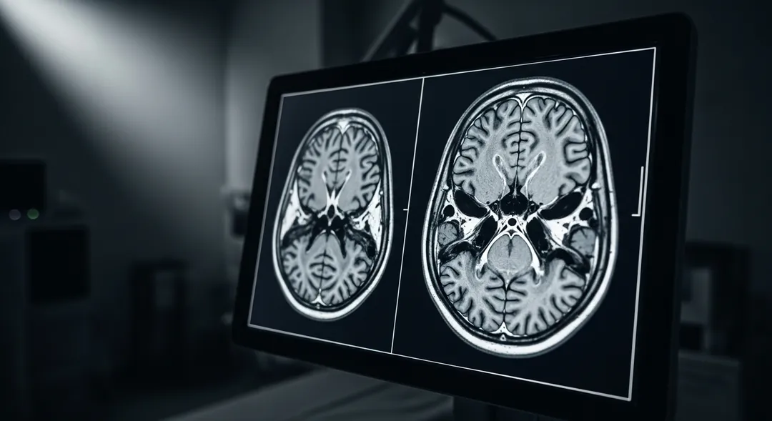 RBrain MRI · T1/T2/FLAIR · AI-generated imageAI-generated image · demonstration
Brain MRI with T1, T2, FLAIR, DWI, ADC and post-contrast T1 sequences. A large complex growth is visible in the left temporal area with surrounding swelling causing a midline shift. The AI identified 6 specific observations.
Axial MRI brain with T1, T2, FLAIR, DWI, ADC and post-contrast T1 sequences. Images are of diagnostic quality.
Want this for your own MRI?
Demonstration only, not a diagnosis. Your report describes the images you upload.
Learn about MRI and how the analysis works
What is magnetic resonance imaging (MRI)?
MRI uses a magnetic field, radio waves and a computer, not X-rays, to create detailed images of the inside of the body.
What the examination shows depends on the scan itself. This tool does not perform or repeat the examination; it only explains the images you upload.
How sequences differ — further down this pageHow does AI analysis of MRI images work?
One analysis is one submission of up to 10 images, read together; they can come from one series or several. From a DICOM folder, the tool first pre-selects evenly spaced images, and you can change the selection.
The AI (Google's general-purpose Gemini model) receives only the pixels of those images and the details you enter: age, sex, notes and an optional attachment. It does not receive the DICOM metadata, the other slices or a 3D volume.
You get a written report: a plain-language explanation next to clinical-style sections (findings, impression, next steps). It is an AI explanation to help you understand the images and prepare questions, not a diagnosis or a radiologist's report.
See the demonstration reportWhat you can do with your MRI images
Analyse selected images
Up to 10 images in one analysis, read together. Images you do not select are not analysed.
A report in clinical and plain language
A plain-language explanation next to clinical-style sections, as PDF or Word. It is not a diagnosis.
See the demonstration reportAsk about your result
Ask follow-up questions about your full report. The chat does not change the report or replace a doctor.
Combine images from several sequences
Mix images from different series in one analysis. The tool does not recognise series by itself, so note them in the form if you know them.
How sequences differOnline DICOM viewer
Look through .dcm files in your browser before choosing images: scroll, zoom, brightness and contrast.
Open the DICOM viewerHow your data is processed and deleted
Your images and the details you enter are processed by Google's Gemini API, an external processor. One image per analysis is stored for 30 days.
Read the privacy policyInformation by body area
Each body area is scanned with its own set of sequences and has its own report vocabulary. Start here if you know which part was scanned.
One study, several sequences
KNEE_MR_2026 — 6 series, 312 images
Fluid is dark on T1 and bright on T2. A finding visible on one sequence can be invisible on the next. Which series a sentence in the report refers to changes what that sentence means.
Words that only appear on an MRI report
- T1 / T2 weighting
- Two ways of timing the measurement. Fat is bright on T1; fluid is bright on T2. The same tissue, two different pictures.
- FLAIR
- A T2 image with the signal from free fluid suppressed, so changes next to fluid-filled spaces stand out. Common in brain studies.
- DWI / ADC
- Diffusion imaging and the map computed from it. They are read as a pair — bright on DWI means little on its own.
- Fat saturation (fat-sat, STIR)
- Fat signal deliberately removed so that swelling and oedema in bone and soft tissue become visible.
- Gadolinium
- MRI contrast agent. Post-contrast series are labelled separately; comparing them to the pre-contrast series is the point.
- Motion artifact
- Smearing from movement during a sequence that can take several minutes. Frequently noted in the report itself.
What an MRI does not show
Stated up front, because it bounds anything read from the images.
Cortical bone and fresh calcification are poorly seen. CT is the study that answers those questions.
A sequence that was not acquired cannot be inferred. If the exam has no post-contrast series, no reading adds one.
Metal implants. They can cause artefacts and make parts of the images harder to assess.
One exported image is one sequence at one slice. A phone photo of a screen carries less again.
Frequently asked questions
What MRI file formats can I upload?
A DICOM (.dcm) series as a folder or multiple files, or individual MRI images already exported as JPG or PNG.
How many images from my MRI are analysed?
Up to 10 per analysis. One analysis is one submission of up to 10 images, read together, from one series or several; images you do not select are not analysed. From a DICOM folder the tool pre-selects evenly spaced images, and you can change the selection. To have more images explained, start another analysis.
Do I need an account, and what is free?
Yes, a free account is required before the analysis runs. A new account (one per email address) gets one free analysis with the full report, including follow-up questions in the chat. Further analyses are paid.
What do T1 and T2 mean on my MRI report?
T1 and T2 are two common ways an MRI machine can capture the same tissue. T1-weighted images show fat and anatomy clearly; T2-weighted images make fluid appear bright, which helps highlight swelling or inflammation.
Why does my MRI report mention ‘sequences’?
A sequence is one specific scanning technique used during your MRI (for example, T1, T2, or FLAIR). A single MRI study usually combines several sequences to show different types of tissue detail.
Can I upload an MRI without contrast?
Yes. Contrast (gadolinium) is not required. The AI receives only the pixels of the images you select and the details you enter (age, sex, notes, an optional attachment), not the file's technical data (DICOM metadata). It can therefore say whether contrast was used only if the images themselves or your notes show it, for example if you write which series is post-contrast.
What will my MRI analysis report include?
Your MRI analysis report includes a Simplified Explanation in plain language, plus clinical Findings, Impression, and Next Steps & Recommendations.
How long do you keep my uploaded scan?
Uploaded images are deleted 30 days after upload. Deleted files may remain in a recoverable backup for up to 7 more days. Original DICOM files, additional images and attachments are not stored by us: only one image per analysis is kept, for those 30 days. Your report and chat stay in your account until you delete them.
Medical disclaimer
X-Ray AI Analyzer is an AI-powered tool for informational purposes only. It is not a certified medical device. AI analysis must always be reviewed by a qualified healthcare professional.