Free AI CT Scan Analysis — understand your images
Select up to 10 images from your CT files and get:
- A plain-language explanation of the selected images
- A structured summary of the observations — add symptoms or an earlier report for context
- Answers in chat to questions about your report — free account
Thousands
of CT scans analyzed
NPS 88
from CT users
140+
countries
NPS (Net Promoter Score) shows how likely users are to recommend us, on a scale from −100 to 100.
Your scan stays private
- Encrypted in transit & at rest
- One image per analysis kept, deleted 30 days after upload
- Your report stays in your account
What is a CT scan?
A CT (computed tomography) scan combines many X-ray measurements into thin cross-sectional slices of your body. One study usually contains hundreds of slices, saved as DICOM files, that together form a 3D picture. CT is especially good at showing bones, lungs, blood vessels and abdominal organs.
How the analysis works
You choose up to 10 images: from a DICOM folder the app pre-selects evenly spaced images, and you can change the selection before you confirm it. The AI reads only the confirmed images, together with the patient’s age, your notes and any earlier report you attach, and explains what it sees in plain language. Images you don’t select are not analyzed. It is not a diagnosis and does not replace a radiologist.
What your report includes
A plain-language explanation and a structured summary of what the AI sees in the images you select — so you can prepare questions for your next appointment.
Scan type and image quality
The likely body area and whether the images are clear enough to explain. The AI cannot confirm whether contrast was used or in which phase — add that in the notes or attach the earlier report.
Organs and anatomy
An explanation of the organs and structures visible in the images you upload — for example lungs, brain, liver, kidneys or spine.
Observations on your images
What the AI sees in the selected images, with where it is and how certain the description is. The AI can miss abnormalities, and not every feature can be identified.
Plain-language explanations
The observations in your AI report are explained in everyday words, so terms like “nodule” or “contrast phase” make sense.
Free CT scan viewer
Open your DICOM files in the browser and scroll through the slices — no software to install.
Open the viewer →Privacy first
Your scans are encrypted in transit and at rest. We keep one image from each analysis so you can see it with your report. It is deleted automatically 30 days after upload. Original DICOM files are never stored.
Example report (X-ray)
This demonstration uses a chest X-ray. A CT report has the same structure: findings, impression and next steps, each explained in plain language.
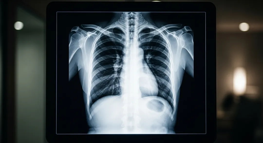 RChest X-Ray · PA · AI-generated imageDemonstration
This is a chest X-ray, PA view. The lungs are mostly clear with mild increased markings in the left lower area. Heart within normal size. The AI identified 4 specific observations in the full report.
PA chest radiograph of diagnostic quality. No prior studies available for comparison.
Want this for your own CT?
Demonstration only, not a diagnosis. Your report describes the images you upload.
What CT users say
“Very thorough and clear explanation.”
“The report included the specific red flag areas and possible factors causing them (e.g. shadows, appearance of bone loss).”
“Very well analysis. It encourages us to research the whole anatomical region, the need for further imaging, and preparation for specialist consultation. It also allows for follow-up questions to the AI Assistant.”
Common terms in CT reports
- Hounsfield units (HU)
- The density scale used to describe tissue on CT.
- Window / level
- Display settings that bring out lung, soft tissue or bone.
- Contrast phase
- When the images were taken after contrast was injected.
- Slice thickness
- How thin each image is — thinner slices show smaller details.
- MPR (multiplanar reconstruction)
- The same scan rebuilt from the front or the side, not just top-down.
- Streak artifact
- Bright or dark lines caused by metal or movement, not by disease.
What a CT image cannot show
- How organs workCT shows structure, not function — for example heart rhythm or hormone levels.
- Fine soft-tissue detailLigaments, cartilage, nerves and small brain changes are usually clearer on MRI.
- Very early changesTiny lesions or early inflammation may not be visible yet.
- The cause of every symptomPain or tiredness can have causes that don’t show on any scan.
What this analysis cannot do
- Images you did not selectOnly the confirmed images (up to 10) are analyzed — the rest of the study is not.
- What a finding meansAn image alone may not establish what a finding means or whether it is benign. Your clinician decides whether further assessment is needed.
- That nothing is wrongAn AI report with no abnormal findings does not rule out disease.
- A diagnosisThat comes from your doctor, combining the scan with your history and tests.
Frequently asked questions
Do I need to upload every slice from my CT scan?
Select the files from your CT folder. From DICOM files, the app pre-selects up to 10 evenly spaced images, and you can change the selection before the analysis. The report covers only the confirmed images. If you only have a single image, you can upload just that.
What CT file formats can I upload?
DICOM files from the folder on your CD or USB — often ending in .dcm or with no extension at all — or JPG and PNG images.
Can I add symptoms, notes or an earlier report?
Yes, if you want to. The patient’s age is required; symptoms, notes and one attached file — for example an earlier report, as an image or PDF — are optional. The AI reads them together with the selected images.
Is my first CT report really free?
Yes. Your first full CT report is free and no credit card is needed. After that, analyses start from $1.75 each in the Plus package — see pricing for all packages.
How long do you keep my uploaded scan?
We keep one image from each analysis so you can see it with your report. It is deleted automatically 30 days after upload. Original DICOM files are never stored. Your report stays in your account, and you can download it as PDF or Word.
What does “with contrast” or “non-contrast” mean on my CT report?
“With contrast” means a dye was injected, usually into a vein, so blood vessels and organs stand out more clearly. “Non-contrast” means no dye was used — common for head injuries, kidney stones or bone problems. Your report states which type of scan you had.
Can I ask questions about my report?
Yes. With a free account you can ask the AI follow-up questions about your own report — for example what a term means or what to ask your doctor.
Can AI replace my radiologist?
No. The AI explains the images you upload to help you understand them and prepare for your appointment. A report with no abnormal findings does not rule out disease. Only a qualified doctor can make a diagnosis, taking into account your symptoms, history and other tests.
CT analysis by body area
 COMING SOON
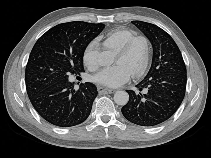
Chest CT
Lungs, nodules, pleura
 COMING SOON
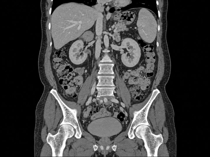
Abdomen & pelvis CT
Liver, kidneys, bowel
 COMING SOON
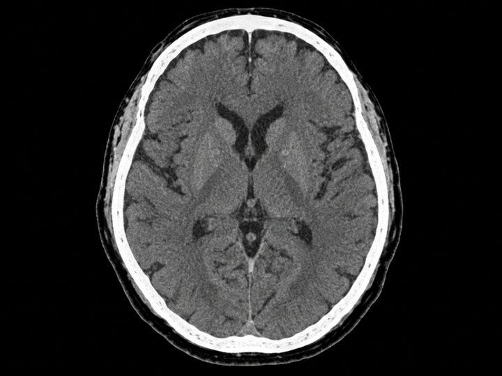
Head CT
Brain, sinuses
 COMING SOON
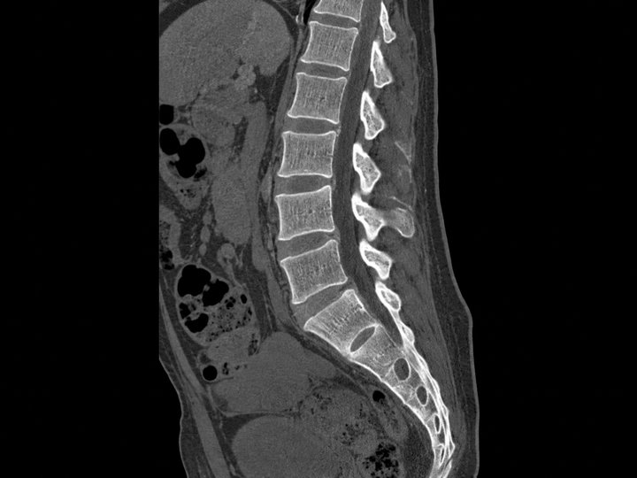
Spine CT
Cervical, lumbar
 COMING SOON
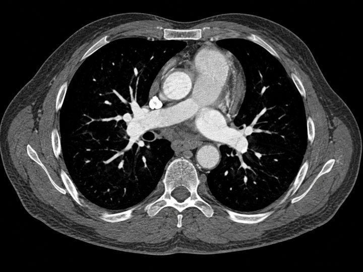
CT angiography
Vessels, contrast phases
Guides about CT scans
CT with vs without contrast: what changes
How to export DICOM files from a hospital CD
Hounsfield units explained
Ready to understand your CT scan?
Select your CT files and get a plain-language explanation with a structured summary. No credit card for your first report.
Upload your CT scanMedical disclaimer
X-Ray AI Analyzer provides AI-generated reports for informational and educational purposes only. It is not a certified medical device and does not provide a medical diagnosis, treatment or medical advice.
AI reports can be incomplete or wrong, and a report with no abnormal findings does not rule out disease. Every report must be reviewed by a qualified healthcare professional, who will interpret it together with your symptoms, medical history and other tests. Never ignore professional medical advice or delay seeking it because of something you read in a report.
If you think you may have a medical emergency, call your local emergency number or go to the nearest emergency department right away.